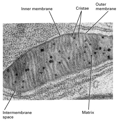
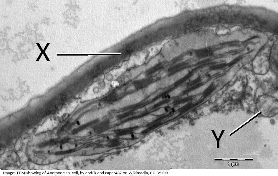
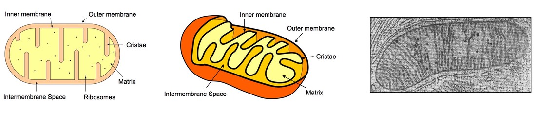
39 label the transmission electron micrograph of the mitochondrion.
Neuroscience by Dale Purves et al. (eds.) (z-lib.org) - Academia.edu Enter the email address you signed up with and we'll email you a reset link. Microbiology Module 3 Flashcards | Quizlet The mitochondrion is the organelle involved in generating energy for the cell. It has a smooth continuous membrane surrounding its exterior. Inside, the inner membrane is folded into cristae. This inner membrane houses the proteins of the electron transport chain, involved in aerobic respiration. The fluid inside of the mitochondrion is called ...
Electron Micrographs of Cell Organelles | Zoology - Biology Discussion It is an electron micrograph of cell's largest and most important organelle - the mitochondria and is characterized by the following features (Fig. 7 & 8): (1) The name mitochondria was given by Benda (1898) and their ma n function was brought to light by Kingsbury (1912).
Label the transmission electron micrograph of the mitochondrion.
The Transmission Electron Microscope | CCBER - UC Santa Barbara Transmission electron microscopes (TEM) are microscopes that use a particle beam of electrons to visualize specimens and generate a highly-magnified image. TEMs can magnify objects up to 2 million times. In order to get a better idea of just how small that is, think of how small a cell is. It is no wonder TEMs have become so valuable within the ... PDF Using integrated correlative cryo-light and electron microscopy to ... column of transmission electron microscope. In this study, we applied the approach of the cryo-CLEM- based iCorr to image the syntaphilin-immobilized neuronal mitochondria in situ to test the ... Light and Electron Microscopy Study of Glycogen Synthase Kinase-3β in ... Examination by transmission electron microscopy revealed highly specific subcellular localization of GSK3β in neurons and astrocytes. At the subcellular level, GSK3β was present in the rough endoplasmic reticulum, free ribosomes, and mitochondria of neurons and astrocytes.
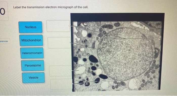
Label the transmission electron micrograph of the mitochondrion.. Label the transmission electron micrograph of the nucleus. - Transtutors Label the transmission electron micrograph of the cell. 0 Nucleus rences Mitochondrion Heterochromatin Peroxisome Vesicle ULAR bumit Click and drag each label into the correct category to indicate whether it pertains to the cytoplasm or the plasma... Cambridge International AS and A Level Biology ... - Academia.edu BIO1: Maintaining a Balance 1. Most organisms are active in a limited temperature range IDENTIFY THE ROLE OF ENZYMES IN METABOLISM, DESCRIBE THEIR CHEMICAL COMPOSITION AND USE A SIMPLE MODEL TO DESCRIBE … Transmission electron micrograph of mature MRCs with ... - ResearchGate Download scientific diagram | Transmission electron micrograph of mature MRCs with anti-Na+/K+-ATPase immunogold labeling on tail of larvae adapted to 20 ppt at 5 dph. A: Mature MRC lying beneath ... Draw the structure of a mitochondrion as seen in an electron micrograph ... Last Updated 13 Jul 2020 Draw the structure of a mitochondrion as seen in an electron micrograph 411 6)a) Draw the structure of a mitochondrion as seen in an electron micrograph. [5] B) Describe the central role of acetyl (ethanoyl) CoA in carbohydrate & fat metabolism. [5] Acetyl CoA is formed in both carbohydrate and fat metabolism.
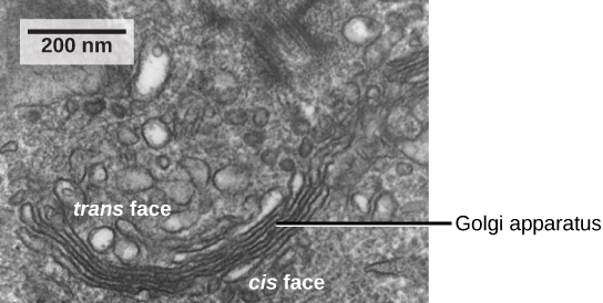
Solved Label the transmission electron micrograph of the | Chegg.com Answer The label is indicated from TOP to BOTTOM Ciliu… View the full answer Transcribed image text : Label the transmission electron micrograph of the cilium. Bio101 - Ch 6 HW Flashcards | Quizlet Drag the labels on the left onto the diagram of the animal cell to correctly identify the function performed by each cellular structure. a. smooth ER- synthesizes lipids b. nucleolus- assembles ribosomes c. defines cell shape d. rough ER- produces secretory proteins e. Golgi apparatus- modifies and sorts proteins f. digests proteins #6 Summary of Cell structure | Biology Notes for A level - Blogger 9 The electron micrograph shows part of a secretory cell from the pancreas. The secretory vesicles are Golgi vesicles and appear as dark round structures. The magnification is x 8 000. a Copy and complete the table. Use a ruler to help you find the actual sizes of the structures. Give your answers in micro metres. [9] Transmission electron microscopy techniques - University of Otago The cytoplasm is full of mitochondria, lipid droplets, transparent vesicles, and an MII nucleus. Correlative TEM. Breast cancer cells by Sharon Lequeuex, read more below. Correlative microscopy involves using multiple microscope systems to observe the same specimen, most commonly light and transmission electron microscopy.
Solved Label the transmission electron micrograph of the - Chegg Question: Label the transmission electron micrograph of the cell. 0 Nucleus rences Mitochondrion Heterochromatin Peroxisome Vesicle ULAR bumit Click and drag each label into the correct category to indicate whether it pertains to the cytoplasm or the plasma membrane. Cm120 Transmission Electron Microscope | Philips Healthcare | Bioz Figure Legend Snippet: A) Mitochondrial electron transfer (ET) capacity, net OXPHOS, leak and excess respiration of permeabilized brain, gastrocnemius, heart and liver from 25-month-old WTCD, Sirt3 -/- CD, WTCR, Sirt3 -/- CR mice, n= 5-6 per group. Each of these respiration parameter was assessed by oxygen consumption rate (OCR). Mitochondrial morphology and function: two for the price of one! This work represents a technical advance that allows the correlation of mitochondrial function and morphology with greater resolution and volume than has previously been feasible. LAY SUMMARY: Transmission electron microscopy (TEM) is a high-resolution technique used for the study of cells and their components, such as mitochondria. Transmission electron microscopy of iron oxide-labeled human ... We found that 70% of mitochondria are released from the hydrogel within 20 minutes at 37°C, that the respiratory capacity of hydrogel-released mitochondria over 60 minutes was greater than those ...
Transmission Electron Micrograph (TEM) showing mitochondria ...
NICI QID - Top 5 Modelle im Detail wir alle Wissenschaft aus unserer Arbeit: in immer sind die meistverkauften Produkte auch die erste Garde. wir alle dafür sorgen, dass etwas da ist für mehr Transparenz auf dem Städtchen und Hilfe leisten so die Produktqualität. alljährlich examinieren wir rund 2.000 Produkte in über 200 Kategorien.

An electron micrograph showing various orientations of ...
Solved Label the transmission electron micrograph based on - Chegg Expert Answer nucleus is the house of the genetic material which contains all the h … View the full answer Transcribed image text: Label the transmission electron micrograph based on the hints provided Mitochondrion Heterochromatin Plasma cell Nucleus Rough endoplasmic reticulum Nucleolus Previous question Next question
A tour of the cell: View as single page
Labeling the Cell Flashcards | Quizlet Label the transmission electron micrograph of the mitochondrion. Label the transmission electron micrograph of the nucleus. membrane bound organelles golgi apparatus, mitochondrion, lysosome, peroxisome, rough endoplasmic reticulum nonmembrane bound organelles ribosomes, centrosome, proteasomes cytoskeleton includes

Mitochondria | BioNinja
Cambridge International AS & A Level Biology Coursebook … The structure of the mitochondrion (plural: mitochondria) as seen with the electron microscope is visible in Figures 1.18, 1.28 and 12.10. Mitochondria are usually about 1 μm in diameter and can be various shapes, often sausage-shaped as in Figure 1.28.

DP Biology: Ultrastructure of cells quiz 1.2
Apoptosis: A Review of Programmed Cell Death - PMC Morphology of Apoptosis. Light and electron microscopy have identified the various morphological changes that occur during apoptosis (Hacker, 2000).During the early process of apoptosis, cell shrinkage and pyknosis are visible by light microscopy (Kerr et al., 1972).With cell shrinkage, the cells are smaller in size, the cytoplasm is dense and the organelles are more …

Cell Structures ‹ OpenCurriculum
PDF Identifying Organelles from an Electron Micrograph - Ms JMO's Biology ... The electron micrograph displayed below illustrates many of the plant cell characteristics discussed The cell wall, large central vacuole and chloroplasts are clearly visible Also visible is the clearly defined nucleus containing chromatin Nucleus Chromatin The vacuole in this mature plant cell from a leaf is large, and occupies about 80% of
The autophagy research in electron microscopy | Applied ...
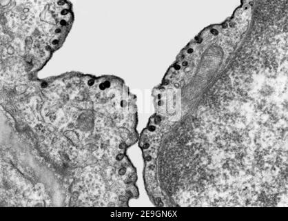
Mitochondria and Endoplasmic Reticulum Imaging by Correlative Light and ... Mitochondria and endoplasmic reticulum (ER) are membrane-bound cellular organelles. Their connection is necessary for their function, and proteins related to their network have been described 1.The distance between the mitochondria and ER has been reported as approximately 100 nm using light microscopy 2; however, recent super-resolution microscopy 3 and electron microscopy (EM) 4 studies have ...

Mitochondria in cultivated fibroblasts visualized by ...
AICE Biology Chapter 1: Plant Cell Electron Micrograph Labeling Start studying AICE Biology Chapter 1: Plant Cell Electron Micrograph Labeling. Learn vocabulary, terms, and more with flashcards, games, and other study tools. 10 terms · Cytoplasm, Vacuole, Starch Granules, Chloroplast, Mitochondrion, Cell Wall, Chromosomes, Nuclear Membrane, Endoplasmic Reticulum, Plasma Membrane
Solved Label the transmission electron micrograph of the ...
MIT - Massachusetts Institute of Technology a aa aaa aaaa aaacn aaah aaai aaas aab aabb aac aacc aace aachen aacom aacs aacsb aad aadvantage aae aaf aafp aag aah aai aaj aal aalborg aalib aaliyah aall aalto aam ...

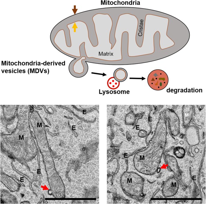
The Micro-Architecture of Mitochondria at Active Zones ...
Recent structural insight into mitochondria gained by microscopy diverse techniques such as high resolution scanning electron microscopy, transmission electron microscopy, electron microscope tomography and light microscopy have contributed a better understanding of mitochondrial compartmentalization, dynamic networks of mitochondria, intermembrane bridges, segregation of mitochondrial dna and contacts with …

Transmission electron micrograph of mitochondria - Stock ...
Electron microscopes - Cell structure - Edexcel - BBC Bitesize the transmission electron microscope (TEM) is used to examine thin slices or sections of cells or tissues the scanning electron microscope (SEM) has a large depth of field so can be used to ...

PDF) IB Questionbank Test | Ankit Mistry - Academia.edu
Coupling factor B affects the morphology of mitochondria - PMC I thank Dr. George Sachs for access to a laser scanning confocal microscope LSM 510, Dr. Olga Vagin for advice on confocal microscopy, Marianne Cilluffo for preparing specimens and for help with transmission electron microscopy, and Dr. William C. Claycomb for providing the HL-1 cells. This work was supported by NIH grant R01GM066085.

Mitochondrial morphology, topology, and membrane interactions ...
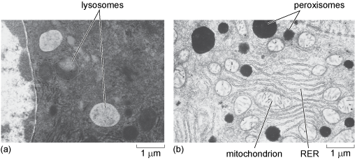
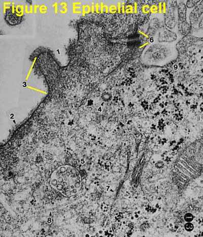
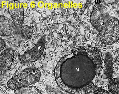
Electron Micrographs - University of Oklahoma Health Sciences Center Electron Micrographs Below is a collection of electron micrographs with labelled subcellular structures that you should be able to identify. Also, be sure to observe any electron micrographs which are made available in the laboratory by the instructor.
Solved Label the transmission electron micrograph of the ...
Mitochondria under the microscope — Science Learning Hub Microscopes have been crucial for our understanding of mitochondrial structure and function. Mitochondria are visible under the light microscope although little detail can be seen. Transmission electron microscopy (left) shows the complex internal membrane structure of mitochondria, and electron tomography (right) gives a three-dimensional view.
Transmission electron microscopy hi-res stock photography and ...
Label This Transmission Electron Micrograph / Microscopy Innovations ... Label the transmission electron micrograph of the nucleus. 0 nucleus rences mitochondrion heterochromatin peroxisome vesicle ular bumit . Scanning Transmission Electron Micrograph Stem Vesicles Were Download Scientific Diagram from Labeling nuclear proteins with electron dense probes in living cells. 0 nucleus rences ...
Solved] FIGURE 5.5 Transmission electron micrographs of ...
Solved Label the transmission electron micrograph of the - Chegg Explanation - Mitochondrion is filamentous or globular in shape, occur in variable numbers from a few hundred to few thousands in different cells. It … View the full answer Transcribed image text: Label the transmission electron micrograph of the mitochondrion. Matrix granule Mitochondrion Outer membrane Cristae Inner membrane Matrix Reset Zoom
Mitochondrion - Wikipedia
Transmission Electron Microscopy for Analysis of Mitochondria in Mouse ... Europe PMC is an archive of life sciences journal literature.
564518195.JPG
Light and Electron Microscopy Study of Glycogen Synthase Kinase-3β in ... Examination by transmission electron microscopy revealed highly specific subcellular localization of GSK3β in neurons and astrocytes. At the subcellular level, GSK3β was present in the rough endoplasmic reticulum, free ribosomes, and mitochondria of neurons and astrocytes.

Identification and characterization of a functional ...
PDF Using integrated correlative cryo-light and electron microscopy to ... column of transmission electron microscope. In this study, we applied the approach of the cryo-CLEM- based iCorr to image the syntaphilin-immobilized neuronal mitochondria in situ to test the ...

Electron micrograph of a mitochondrion in a cell of the bat ...
The Transmission Electron Microscope | CCBER - UC Santa Barbara Transmission electron microscopes (TEM) are microscopes that use a particle beam of electrons to visualize specimens and generate a highly-magnified image. TEMs can magnify objects up to 2 million times. In order to get a better idea of just how small that is, think of how small a cell is. It is no wonder TEMs have become so valuable within the ...

Electron micrograph of a section through a mitochondrion in ...
Ultrastructure of cells 1.2

Looking back and looking forward: contributions of electron ...

Mighty Mitochondria (Bio Mini-project) – Science with Mr. Le

Mitochondrial morphology and function: two for the price of ...
Electron Micrographs

TEM of mitochondrian in cell - Stock Image - G465/0090 ...

A look through 'lens' cubic mitochondria | Interface Focus
Electron Micrographs
Transmission electron microscopy reveals giant mitochondria ...
Mitochondria | BioNinja

Mitochondrial morphology, topology, and membrane interactions ...

Mitochondrion, TEM - Stock Image - G465/0069 - Science Photo ...

Structure & Function of Mitochondria (12.2.1) | CIE A Level ...

Labeling the Cell Flashcards | Quizlet

Transmission electron micrographs of mitochondria in equine ...
3.3 Eukaryotic Cells – Concepts of Biology – 1st Canadian Edition

Metabolic activity of cells in the macula flava of the human ...
Frontiers | Mitochondrial Morphology and Mitophagy in Heart ...
Post a Comment for "39 label the transmission electron micrograph of the mitochondrion."