45 how to label gel electrophoresis images
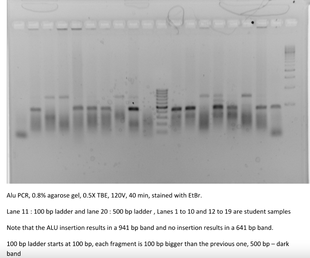
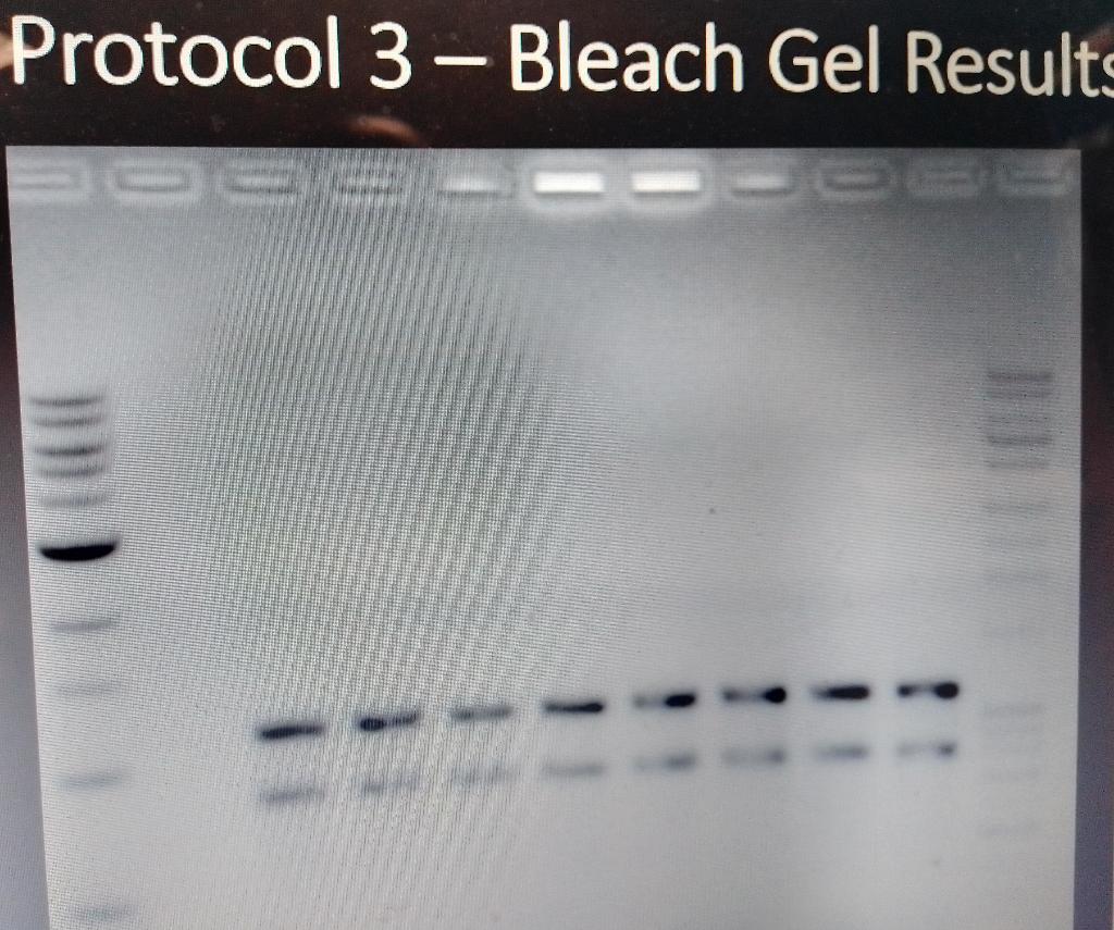
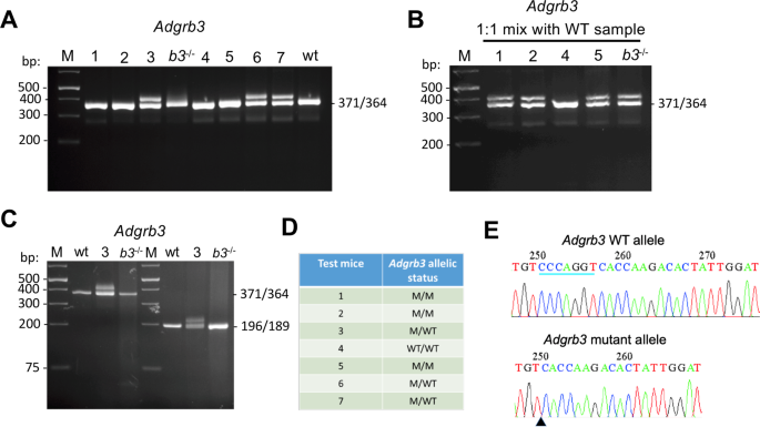
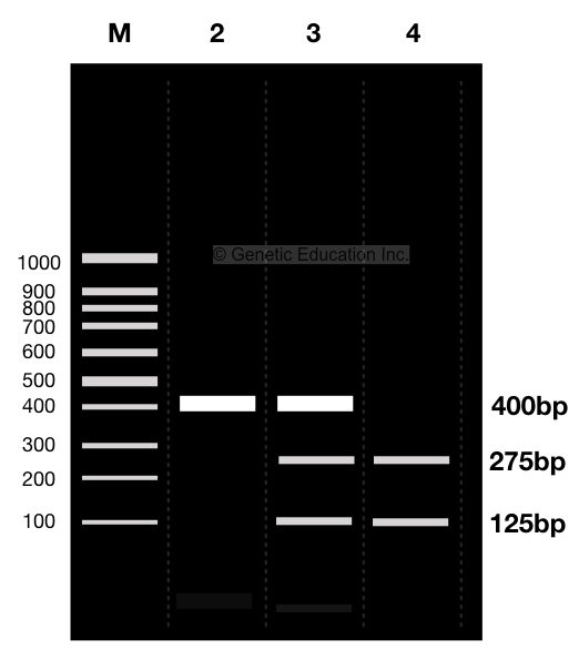
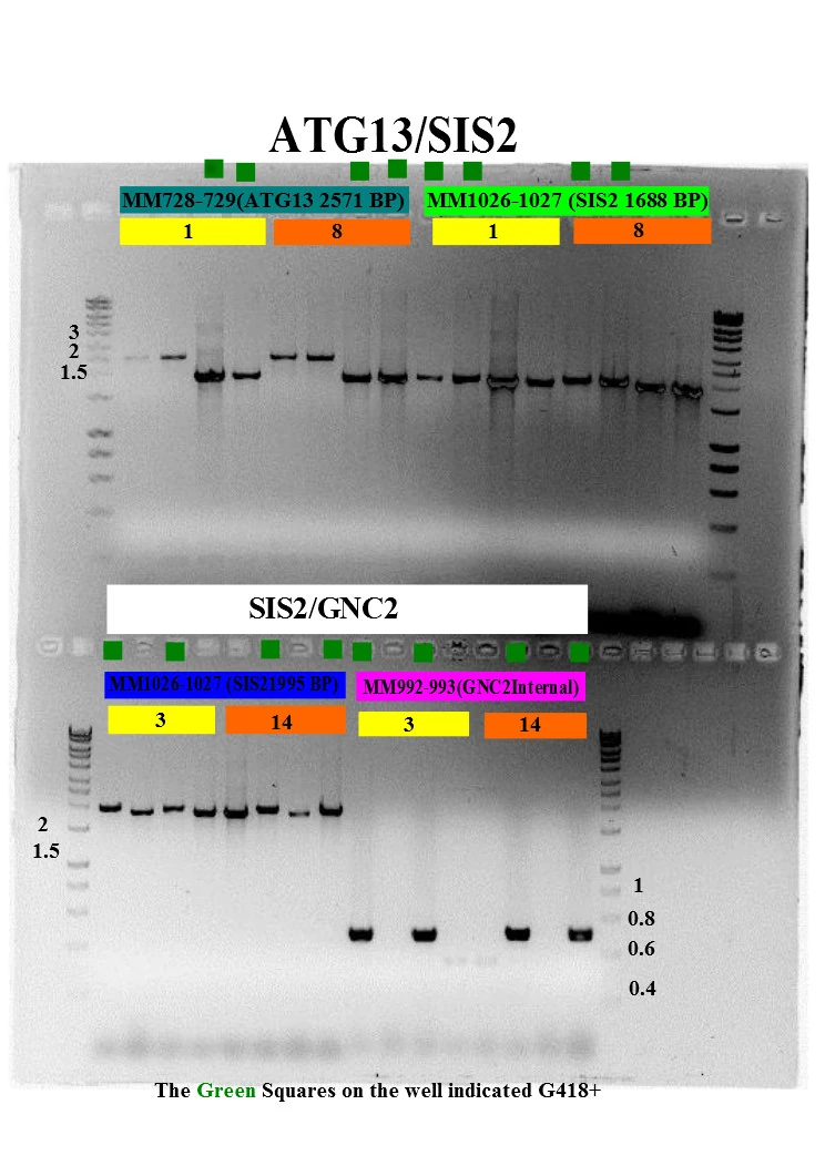
Part 2: Analyzing and Interpreting (Agarose) Gel Electrophoresis Results Now let see some real images. The gel image above is the result of restriction digestion. Lane 3, 5, 7, and 8 are a homozygous normal allele with a 184bp band here one band of 68bp is also present, but it is not visible. Lane 2 is a mutant uncut allele of 252bp. Lane 1 and 6 are heterozygous contain three alleles: 252bp, 184bp and 68bp. PDF 8/13/2009 Tutorial ImageJ Using ImageJ to Quantify Gel Images of interest go to Image/Crop to crop the selection.See below for screenshots. Enhancing the Gel Image This is a typical step when dealing with gel images. You need to adjust the histogram of the image. Please make sure not to blow-out (saturate) the whites. You want to make sure your image has enough dynamic range. Talk to me if you're confused.
ImageJ for Editing & Labelling PCR Gel Image | Biotechnology This Tutorial is all about how to quickly Edit & Label PCR Gel Image Using ImageJ software. Presented by - Elvis SamuelJoin Our Telegram Channel for free Sof...
How to label gel electrophoresis images
Analyzing gels and western blots with ImageJ - lukemiller.org After drawing the rectangle over your first lane, press the 1 (Command + 1 on Mac) key (Command + 1 on Mac) or go to Analyze>Gels>Select First Lane to set the rectangle in place. The 1st lane will now be highlighted and have a 1 in the middle of it. 5. PDF Gel Electrophoresis Size Marker - dia-m.ru The different gel formats for agarose and polyacrylamide gel electrophoresis and the varying sensitivity of staining or detec-tion mean that it is only possible to give an approximation of the recommended DNA amount to be loaded. Most DNA mar-kers show the best separation with loading amounts of 0.5 - 1 µg on agarose gels. Analysis of protein gels (SDS-PAGE) - Rice University The resources on protein gel analysis focus on "routine" gels that are use to separate polypeptides from samples containing a mix of proteins. Such gels are most often stained with Coomassie blue dye, although the principles described here also apply to gels stained by other means. Before starting an analysis one's goals should be defined.
How to label gel electrophoresis images. GelAnalyzer GelAnalyzer 19.1 Analyze gel images from any source Use your digital camera, smartphone, or gel doc system to obtain images. GelAnalyzer will take care of the rest. Automatic lane and band detection With full manual control over adding, modifying, and deleting lanes and bands. Fix run distortions through Rf calibration How to Interpret DNA Gel Electrophoresis Results - GoldBio During gel electrophoresis, you may have to load uncut plasmid DNA, digested DNA fragment, PCR product, and probably genomic DNA that you use as a PCR template into the wells. Your digested DNA fragment is a digested PCR product. The next step is to identify those bands to figure out which one to cut. Gel Electrophoresis. Lane 1: DNA Ladder. How to quantify each band in gel electrophoresis? - ResearchGate you can do an analytical curve in a 1d gel, with known amounts of bsa for example, use photoshop to quantify the pixels (the curve would be pixels x protein mass you applied for each well) and then... How can I modify a photograph of gel electrophoresis taken with ... What exactly you need to modify, if u wanted to increase resolution and size or format u can use photoshop software. This is perfect tool to corred photos and u can also change the format of photo ...
Annotating A Gel | Get Your Science On Wiki | Fandom Part 1. Photo Editing: 1.Take your JPG or PNG file of your Gel and open it with a photo editing program (GIMP). 2. Under "Image" --> "Transform" rotate your picture by 90 degrees so that your wells are on top of the page. 3. Using the Crop tool Cut out the black borders leaving only the gel. 4. Gel Electrophoresis: Basics & Steps - SchoolWorkHelper Basic Steps Aragonese and the buffer are mixed together and microwaved to create the gel. It is poured into a mold and has a "comb" placed in it to make holes for the DNA to be inserted. Once it has cooled the comb is removed. The gel is then placed in the gel electrophoresis box and buffer solution is poured onto it. Agarose gel electrophoresis of DNA - Principle, Protocol and Uses Image 2: An agarose gel electrophoresis is a process useful in various applications including forensic investigation, molecular cloning, and genetic fingerprinting. Picture Source: news-medical.net Applications of agarose gel electrophoresis. It helps identify unknown samples. It is a method of choice for checking the quality and accuracy of other procedures. Gel electrophoresis Images, Stock Photos & Vectors - Shutterstock Find Gel electrophoresis stock images in HD and millions of other royalty-free stock photos, illustrations and vectors in the Shutterstock collection. Thousands of new, high-quality pictures added every day.
Solved Please label the images to review the process of - Chegg Expert Answer Answer) The complete DNA molecules with primer and buffer containing nucleotides and taq polymer … View the full answer Transcribed image text: Please label the images to review the process of polymerase chain reaction and how its products can be analyzed using gel electrophoresis. CHAPTER 10 Flashcards | Quizlet Please label the images to review the process of polymerase chain reaction and how its products can be analyzed using gel electrophoresis. Match the components of a typical PCR reaction with the function they serve. ... Please label the images to review the process of screening bacterial clones for those containing a donor gene. Other sets by ... Addgene: Protocol - How to Run an Agarose Gel If you do not add EtBr to the gel and running buffer, you will need to soak the gel in EtBr solution and then rinse it in water before you can image the gel. Pour the agarose into a gel tray with the well comb in place. *Pro-Tip* Pour slowly to avoid bubbles which will disrupt the gel. Solved Please label the images to review the process of - Chegg Question: Please label the images to review the process of polymerase chain reaction and how its products can be analyzed using gel electrophoresis. Dam Deration Denaturation 1 се DNA Replication Pricing Olgorde sha and of of arcon A Cole 770 Restriction andonucleases selectively cleaving sites of DNA cony Piring w Opelweg () Restriction ...
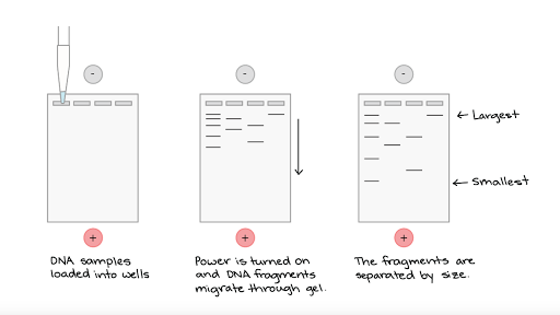
Gel electrophoresis (article) - Khan Academy When a gel is stained with a DNA-binding dye and placed under UV light, the DNA fragments will glow, allowing us to see the DNA present at different locations along the length of the gel. The bp next to each number in the ladder indicates how many base pairs long the DNA fragment is. A well-defined "line" of DNA on a gel is called a band.
Gel Electrophoresis: Definition, Principle, and Application Electrophoresis is a process used for the separation of macro and micro molecules in an electric field by applying charges at both the extents. The mixture of substances is spread in the supporting film. The supporting films are placed in a salt solution filled in a container, where one container holds a cathode and the other carries an anode.
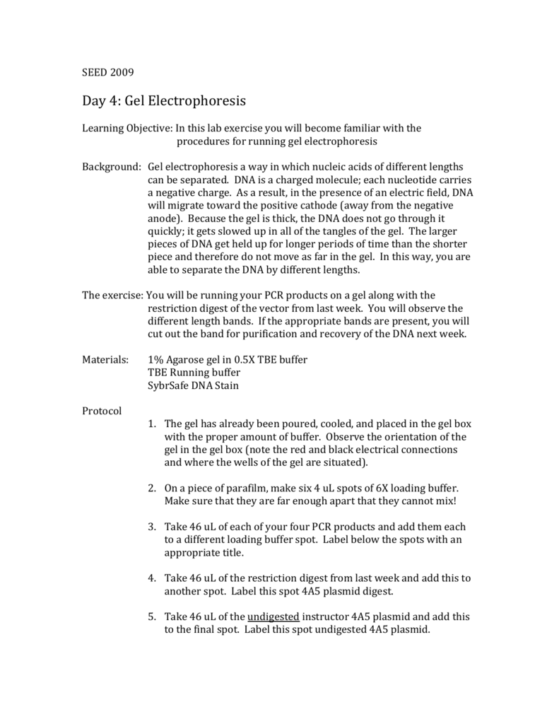
PDF Lab 4: Gel Electrophoresis - Vanderbilt University Gel electrophoresis Gel electrophoresis is a method of separating DNA fragments by movement through a Jello-like substance called agarose. Derived from a seaweed polysaccharide, agarose gels form small pores that act as sieves to separate DNA based on size; whereby smaller DNA molecules move through the pores faster and easier than larger ...
Gel Electrophoresis - Definition, Purpose and Steps - Biology Dictionary The gel chamber wells are loaded with the DNA samples and usually, a DNA ladder is also loaded as reference for sizes.. 6. Electrophoresis. The negative and positive leads are connected to the chamber and to a power supply where the voltage is set. Turning on the power supply sets up the electric field and the negatively charged DNA samples will start to migrate through the gel and away from ...
What is gel electrophoresis? - YourGenome Illustration of DNA electrophoresis equipment used to separate DNA fragments by size. A gel sits within a tank of buffer. The DNA samples are placed in wells at one end of the gel and an electrical current passed across the gel. The negatively-charged DNA moves towards the postive electrode. Image credit: Genome Research Limited.
Gel Electrophoresis - an overview | ScienceDirect Topics For gel electrophoresis, a DNA sample is loaded at one end of a gel matrix (usually agarose or acrylamide) that provides a uniform pore size through which the DNA molecules can move. Application of a constant electric field causes DNA fragments (all have a uniform, strong negative charge) to migrate toward the cathode.
A Complete Guide for Analysing and Interpreting Gel Electrophoresis Results let see some of the gel images of PCR fragments. 2% gel is required to separate PCR products because PCR products are the smaller fragments of DNA nearly ~100bp to ~1500bp. Image 1: The image is captured under the UV transilluminator instead of the gel doc system to show you the effect of EtBr on the gel electrophoresis results.
3 Ways to Read Gel Electrophoresis Bands - wikiHow Hold a UV light up to the gel sheet to reveal results when using a UV-based dye. With your gel sheet in front of you, find the switch on a tube of UV light to turn it on. Hold the UV light 8-16 inches (20-41 cm) away from the gel sheet. Illuminate the DNA samples with the UV light to activate the dye and read the results.
InDesign Labeling / Annotating PCR Gel Pictures - Advanced Tutorial ... In this tutorial we will learn how to annotate Agarose Gel Pictures with Adobe InDesign CS5. I see people often labeling pictures in Photoshop and I can't re...
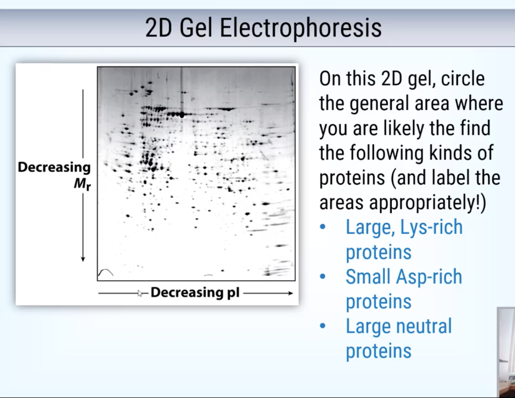
Analysis of protein gels (SDS-PAGE) - Rice University The resources on protein gel analysis focus on "routine" gels that are use to separate polypeptides from samples containing a mix of proteins. Such gels are most often stained with Coomassie blue dye, although the principles described here also apply to gels stained by other means. Before starting an analysis one's goals should be defined.
PDF Gel Electrophoresis Size Marker - dia-m.ru The different gel formats for agarose and polyacrylamide gel electrophoresis and the varying sensitivity of staining or detec-tion mean that it is only possible to give an approximation of the recommended DNA amount to be loaded. Most DNA mar-kers show the best separation with loading amounts of 0.5 - 1 µg on agarose gels.
Analyzing gels and western blots with ImageJ - lukemiller.org After drawing the rectangle over your first lane, press the 1 (Command + 1 on Mac) key (Command + 1 on Mac) or go to Analyze>Gels>Select First Lane to set the rectangle in place. The 1st lane will now be highlighted and have a 1 in the middle of it. 5.



















Post a Comment for "45 how to label gel electrophoresis images"