44 clam labeled diagram
PDF LS - Activity #6 Clam - Diagram - CIRCLE Draw a detailed diagram that shows the structure of a clam. 1. Diagram must be on 8.5 X 11 inch drawing paper. 2. Diagram must take up 80% of the sheet of paper. 3. Labeling must be in black ball point pen 4. Diagram must be colored using colored pencils. 5. This cover sheet must be stapled neatly to your diagram. Clam Dissection - BIOLOGY JUNCTION Answer the questions on your lab report & label the diagrams of the internal structures of the clam. Also, use arrows on the clam diagram to trace the pathway of food as it travels to the clam's stomach. Continue the arrows showing wastes leaving through the anus. CLICK HERE FOR LAB QUESTIONS CLICK HERE FOR PHOTOS/QUIZ
PDF Lab 5: Phylum Mollusca - Amherst clam. These saltwater clams are sedentary and live intertidally and in shallow subtidal areas of sand flats on the east coast of the U.S. The morphology and anatomy of modern bivalves have been much altered from those of ancestral mollusks, which had a distinct anterior end with a mouth and a posterior end with an anus (Figure 1).

Clam labeled diagram
Clam Anatomy Labeling Page - exploringnature.org The style of citing shown here is from the MLA Style Citations (Modern Language Association). When citing a WEBSITE the general format is as follows. Author Last Name, First Name (s). "Title: Subtitle of Part of Web Page, if appropriate." Title: Subtitle: Section of Page if appropriate. Sponsoring/Publishing Agency, If Given. clam anatomy101 | Clam Anatomy Diagram | tanks4thememories | Flickr Clam Anatomy Diagram. This site uses cookies to improve your experience and to help show content that is more relevant to your interests. New Page 1 [ez002.k12.sd.us] The oldest part of the clam shell is the umbo, and it is from the hinge area that the clam extends as it grows. Procedure 1. Put on your lab apron, safety glasses, and plastic gloves. 2. Place a clam in a dissecting tray and identify the anterior and posterior ends of the clam as well as the dorsal, ventral, & lateral surfaces. 3.
Clam labeled diagram. Solved 4 5 7 6 This is a diagram of a dissected clam. - Chegg Biology. Biology questions and answers. 4 5 7 6 This is a diagram of a dissected clam. [Blank #1] To what phylum do these organisms belong? (Blank #2] Name the structure labeled 4. [Blank #3] Name the structure labeled 5. (hint: this one of 2 structures that hold the shells closed when you try to pry them open) [Blank #4] Name the structure ... Clam Diagram Quiz - By dwhite298 - Sporcle Top User Quizzes in Science. Find the Odd One Out: Numbers (Very Hard) 41. Philippines' Native Animals 33. Pennsylvania's Native Animals 24. Correctly Located Animals VI 17. Oregon's Native Animals 14. Label the Skeleton - 13. Trigonometric Exact Values 11. Identify the Leukocytes (White Blood Cells) 10. Clam Diagram Labeled - schematron.org Use your probe to trace the path of food & wastes from the incurrent siphon through the clam to the excurrent siphon. Answer the questions on your lab report & label the diagrams of the internal structures of the clam. Also, use arrows on the clam diagram to trace . PDF Clam Dissection Guideline - Monadnock Regional High School Clam Dissection Guideline BACKGROUND: Clams are bivalves, meaning that they have shells consisting of two halves, or valves.The valves are joined at the top, and the adductor muscles on each side hold the shell closed. If the adductor muscles are relaxed, the shell is pulled open by ligaments located on each side of the umbo.The clam's foot is used to dig down into the
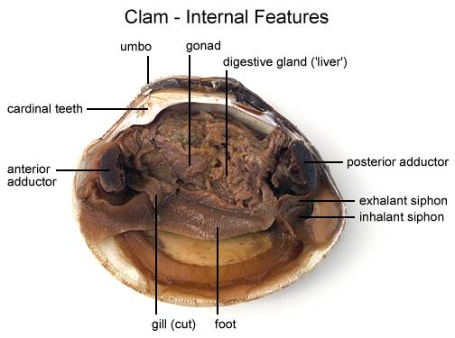
Clam Worm Diagram - schematron.org Alitta succinea (known as the pile worm or clam worm) is a species of marine annelid in the family Nereididae (commonly known as ragworms or sandworms). It has been recorded throughout the North West Atlantic, as well as in the Gulf of Maine and South Africa.Polychaete - WikipediaClam Dissection 6 thoughts on " Clam worm diagram " Iesernamex says: Clam Dissection Lab: Explained | SchoolWorkHelper Answer the questions on your lab report & label the diagrams of the internal structures of the clam. Use arrows on the clam diagram to trace the pathway of food as it travels to the clam's stomach. Continue the arrows showing wastes leaving through the anus. When you have finished dissecting the clam, dispose of the clam as your teacher ... clam anatomy - YouTube This project was created with Explain Everything ™ Interactive Whiteboard for iPad. PDF Taxonomy, Anatomy, and Biology of the Hard Clam Internal Clam 1. Inner surface of left valve 2 Pt dd t l Shell Anatomy Post. adductor muscle 3. Ant. adductor muscle •Hold valves shut 4. Hinges •Ligament holds valves together •Interlocking teeth prevent valves from side slipping when opening and closing 5. Tth Teeth along ventral margin •Prevent valves from sliding when closes 6.
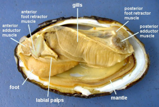
PDF Investigation #5 - Clam Anatomy Locate the following parts of your clam according to the diagram: adductor muscles gills mantle excurrent siphon incurrent siphon stomach mouth foot intestine Lift the gills to find the stomach and intestines. Insert the skewer into the mouth and see that it empties into the stomach. Locate the foot that is used for digging. PDF Diagram Of Clam Anatomy anatomy amp diagram, comprehensive nclex questions most like the nclex, muscle systems types tissue amp facts britannica com, cuttlefish wikipedia, biology dictionary p q macroevolution net, what are the adaptations of mussels answers com, thorax definition and anatomy study com, bivalvia wikipedia Powered by TCPDF ( ) 3 / 3 PDF Diagram Of Clam Anatomy Diagram Of Clam Anatomy Author: OpenSource Subject: Diagram Of Clam Anatomy Keywords: diagram,of,clam,anatomy Created Date: 5/31/2022 4:27:59 AM ... PDF Anatomy of a Clam - University of Florida Obtain clam specimens (fresh or preserved). 2. Divide the class into small groups (2-4 per group when possible). 3. Prepare one dissection kit, pan, and clean-up materials per group. 4. Copy the dissection guide for each student. 5. Copy the Externaland Internal Clam Anatomyhandouts for each student. 6.
Clam Anatomy Diagram | Quizlet Clam Anatomy. STUDY. Learn. Flashcards. Write. Spell. Test. PLAY. Match. Gravity. Created by. Kkelly8267. Terms in this set (12) excurrent siphon. a dorsal tube through which water exits the mantle cavity of a bivalve. incurrent siphon. a ventral tube through which water enters the mantle cavity of a bivalve. posterior adductor muscle...
PDF Biology 11 Name: Clam Dissection 1 anterior posterior 2. Diagram 1: Clam External Anatomy 3. A stick has been inserted between the shells to aid in opening the clam. Place your clam left side up with the dorsal edge away from you. With your blade pointing toward the dorsal edge, slide your scalpel between the upper valve & the top tissue layer.
Clam Diagram & Parts | What Is a Clam? | Study.com Clam Diagram Lesson Summary What Is a Clam? Over 15,000 extant species of clams, ranging in size from a few inches to over six feet in length, inhabit a variety of freshwater and ocean environments...
Clam Anatomy Coloring Page - exploringnature.org Clam Anatomy Coloring Page. PDF for Printing Out. Click Here. Link to More Info About this Animal (with Labeled Body Diagram) Click Here. Citing Research References. When you research information you must cite the reference. Citing for websites is different from citing from books, magazines and periodicals. The style of citing shown here is ...
clam anatomy diagram Quiz - purposegames.com This online quiz is called clam anatomy diagram. This game is part of a tournament. You need to be a group member to play the tournament
clam anatomy Diagram | Quizlet Start studying clam anatomy. Learn vocabulary, terms, and more with flashcards, games, and other study tools.
Clam Dissection - Science by Slim 25. Answer the questions on your lab report & label the diagrams of the internal structures of the clam. Also, use arrows on the clam diagram to trace the pathway of food as it travels to the clam's stomach. Continue the arrows showing wastes leaving through the anus.
PDF Clam Anatomy Exercise Clam Anatomy Exercise . Objective: Students will observe the inside and outside of a clam. Record on the sheet provided. Each student will receive the following materials: fresh clam (in shell), shucked clam, calipers, tray, dissecting tools, clam diagrams . Outside of the clam shell (use live clam):
DOC Clam Dissection - PC\|MAC There are another pair of gills on the right side of the clam. The accompanying diagram shows these parts. You can see the edge of the right mantle below the foot and visceral mass. To see where the heart is located look above the visceral mass above the gills. There is a clear looking region near the top of the clam.


Post a Comment for "44 clam labeled diagram"